Every organism is made of cells. And while most diagnostic procedures take a look at average values across many cells, the ability to analyze a single cell offers great promise in cancer research, stem cell research, and more.
Single Cell Imaging: The Process
Selecting An Imaging Tool
To get the results you need, the right single-cell imaging platform is a must. Advanced platforms let you analyze over 1000 RNA assays in just one experiment. They also offer both visualization and quantification. You can type cells, examine cell-cell interaction, discover biomarkers, determine cell state and function, and more.
Preparing A Sample
Before the imaging itself can occur, you will need to prepare a tissue sample. In most cases, you have two options: formalin-fixed paraffin-embedded (FFPE) or fresh frozen (FF) samples.
The type of sample used depends on your imaging goals. Fresh frozen samples are faster and easier to prepare. They are also ideal for the analysis of molecular genetics as well as DNA and RNA sequencing. That’s because the process of preparing FFPE samples can disrupt molecular data.
On the other hand, FFPE samples have the advantage of not deteriorating rapidly. Once prepared, these samples can be archived at room temperature for decades! Since they can be accessed years later, these samples provide a historical record and are ideal for long-term research.
Isolating A Single Cell
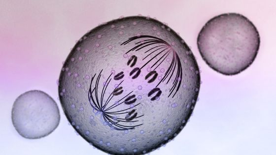
For true single-cell imaging, an imaging platform must first be able to isolate a single cell for analysis. This process is a lot more complex than it appears. An imaging tool identifies cell membrane proteins to accurately determine the boundaries of an individual cell.
Like most advanced technologies, cell segmentation is improving daily. The best platforms use a machine-learning algorithm to constantly develop more accurate ways to focus on a single cell. With these techniques, there is no need to physically separate a single cell from a mass of tissue.
Sample Analysis
One of the advantages of this technique is that it doesn’t require time-consuming sample amplification or tissue expansion. Before the imaging process begins, you just need to fix the targets of your analysis.
From there, the imaging platform will perform a detailed analysis and create an image via repeated hybridization chemistry. Through this process, targeted genes will be illuminated with different dyes. This method is precise enough that it can accurately image even very complex cells like brain cells.
Applying Results
Via an imaging platform, you can both view the analyzed cell and view collected data via a software readout. But how are the results used?
One of the most important applications of single-cell imaging is the identification of spatial biomarkers. By focusing on biomarkers, you can focus on the changes in a single cell’s gene expression based on the treatment of cancer or another disease. And since you can visualize the entire cell, you can see the location of key biomarkers within the cell itself.
This method of analysis also enables you to identify tumor cells in blood before they have extensively multiplied. Similarly, it can help you to discover relatively rare B cells: a must when working to discover antibodies.
Single-cell imaging is also already helping researchers learn more in emerging areas of medicine. Analysis of the complex gut microbiome can help to determine the functions of individual strains of gut bacteria. Since some of these bacteria likely contribute to the development of diseases, this research may help improve the understanding and treatment of various diseases.
Imaging Live Cells in the Body
In some cases, you may want to visualize cells as they work within the body. Bioluminescence is one way to do this. In some studies, researchers have been able to engineer bioluminescent imaging systems to effectively highlight animal cells, even those in deep tissue.
This method has some unique challenges you don’t see with sample-based imaging. However, it allows you to see how individual cells function in vivo, and it’s an important development in the world of single-cell imaging.
Conclusion
Whether you work in oncology, immunology, developmental biology, or a related field, imaging a single cell has the potential to deliver cutting-edge results that can place your practice ahead of the curve. With seemingly endless applications, this technique has the potential to expand the field of medicine as a whole.


